Mohamed A Ghandourah1,
Walied M Alarif1 ![]() ,
Ahmed Abdel-Lateff2,3,
Khalid O Al-Footy4,
Mohamed Halid4,
Sultan S Al-Lihaibi1,
Hajer S Alorfi1
,
Ahmed Abdel-Lateff2,3,
Khalid O Al-Footy4,
Mohamed Halid4,
Sultan S Al-Lihaibi1,
Hajer S Alorfi1
For correspondence:- Walied Alarif Email: wailed1737@yahoo.com
Received: 6 November 2016 Accepted: 20 February 2017 Published: 31 March 2017
Citation: Ghandourah MA, Alarif WM, Abdel-Lateff A, Al-Footy KO, Halid M, Al-Lihaibi SS, et al. Antiproliferative effects of isoprenoids from Sarcophyton glaucum on breast cancer MCF-7 cells. Trop J Pharm Res 2017; 16(3):501-507 doi: 10.4314/tjpr.v16i3.2
© 2017 The authors.
This is an Open Access article that uses a funding model which does not charge readers or their institutions for access and distributed under the terms of the Creative Commons Attribution License (http://creativecommons.org/licenses/by/4.0) and the Budapest Open Access Initiative (http://www.budapestopenaccessinitiative.org/read), which permit unrestricted use, distribution, and reproduction in any medium, provided the original work is properly credited..
Purpose: To evaluate the anticancer activity of isoprenoids of Sarcophyton glaucum on MCF-7 cells and to investigate the potential synergistic effect of doxorubicin.
Methods: Isolation and purification of isoprenoids were performed by applying different planar chromatographic methods (CC and PTLC). Further analyses of the isoprenoids by nuclear magnetic resonance (NMR) and mass spectrometry (MS) carried out to identify the compounds. Sulforhodamine-B (SRB) assay was used to determine the cytotoxic activity of the compounds against the MCF-7 human cell line. Flow cytometric analysis was used to assess their impact on cell cycle of MCF-7. Combination index (CI), when the compounds were combined with doxorubicin, was calculated to determine possible synergism. The isoprenoid compounds were also incubated at ¼ or ½ of their respective half-maximal concentration (IC50) with equimolar concentrations of doxorubicin.
Results: Four known isoprenoid derivatives (1-4) were identified as 10(14)-aromadendrene (1), sarcophinediol (2), ent-deoxysarcophine (3) and sarcotrocheliol acetate (4). It was observed that cells accumulated in pre-G phase as well. CI of compound 3 with doxorubicin was 0.67 and 0.79, respectively, at ¼ and ½ of IC50, indicating overt synergism. This was confirmed by re-assessing the cell cycle stages of MCF-7 cells.
Conclusion: The results indicate that compound 3 exhibits promising cytotoxicity as well as synergism with doxorubicin in MCF-7 cells. This is attributed, at least partly, to its ability to generate intercellular apoptosis induction
Introduction
The marine biota is characterized by ramification of the living organisms which live in the harsh perimeter [1]. Marine invertebrates, particularly soft corals, which lack of natural defenses (e.g., spines) lead to the production of defense metabolites [2].
Alcyonacea or soft corals constitute important organisms of marine invertebrates which are ubiquitous in the tropical sea waters. Moreover, they are considered as a prolific source of unique antiproliferative metabolites [3-5]. For instance, eleutherobin, a tricyclic diterpene displayed high potency in the in vitro induction of tubulin polymerization. This marine natural factor showed specific cytotoxicity toward several cell lines, including those of lung, ovarian, renal and lung [6]. The activity of eleutherobin in cancer cell therapy has been found to be comparable to that of taxol (a very active metabolite isolated from the terrestrial plant) [7].
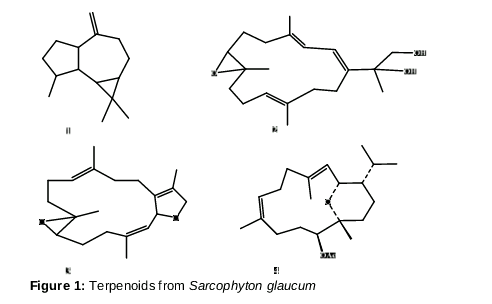
Cancer is a dreadful disease and is the direct cause of almost 14.5 % of all deaths across the world, which is found to increase with the aging of the population [8,9]. Amongst women, breast cancer is the most common type [10]. Anthracycline‑based chemotherapy, particularly, doxorubicin (DOX) is used to treat early stage breast cancer [11]. However, it shows serious adverse effects, including cardiotoxicity [12]. Combining DOX with other cytotoxic agents has been suggested to enhance DOX cytotoxicity and avoid additional toxicity [8]. As combining factors with DOX, metabolites originated from natural sources (marine and terrestrial) and are strongly accepted from scientists and from the public communities, as well [13]. This recent paper further reported the cytotoxicity of isoprenoid derivatives (1-4) isolated from Sarcophyton glaucum () against MCF-7 and the potential synergistic effect of combining those compounds with DOX as well.
Methods
Equipment and reagents
Chromatography: TLC plates (GF 245 Si gel, Merck); PTLC plates (glass supported neutral Al2O3, 20 cm× 20 cm, 25 mm, Merck) and CC (60 G Si gel, Merck). Solvents and Reagents: CDCl3, TMS, 50 % H2SO4/CH3OH, were employed as a solvent for NMR measurements, an NMR internal standard, and a spray reagent, respectively. Doxorubicin, sulforhodamine-B (SRB) and dimethyl sulfoxide (DMSO) are Sigma-Aldrich products. Cell culture materials and fetal bovine serum are Lonza products. Instrument: NMR analysis (AVANCE III WM 600 MHz (1H) and 150 MHz (13C), Bruker). TLC: Thin Layer Chromatography; PTLC: Prep. TLC; CC: Column Chromatography and TMS: Tetramethylsilane.
Collection of S. glaucum
In January 2014, at a depth of 5 - 10 m of North Jeddah Red Sea coast in Saudi Arabia, S. glaucum belonging to Alcyonacea (order; family Alcyoniidae) was collected by SCUBA divers. A voucher specimen (no. SC-2014-10) was kept at Faculty of Pharmacy, KAU, Jeddah, KSA.
Extraction and isolation
In a mixture of dichloromethane miscible with methanol (2:1), 5 kg of S. glaucum was extracted for 24 h (10 L × 3 batches, room temp.). The extract was then filtered, evaporated and extracted under vacuum to obtain a black sticky residue. Re-extraction of the organic residue between water and diethyl ether using a separating funnel followed by drying of the ether layer resulted in getting 30 g ether fraction soluble extract.
The dried residue was physically partitioned on the Si gel column. The elution process was done using n-hexane, followed by a gradual increase in polarity with volumes of Et2O and then replaced volumes of EtOAc.
100 fractions were obtained (F 1-100), the homogeneity and the separation efficiency of the resulted fractions have been examined by investigating the TLC profile using 50 % sulfuric acid reagent or the UV lamp. Purification of fraction F-3 (300.0 mg) eluted with 5 % diethyl ether in n-hexane using prep. TLC with the same elution system (Violet-red zone with H2SO4 reagent, Rf= 0.96, colorless oil, 22 mg, 1). Purification of another one, F-13 (125.0 mg) eluted with 25 % diethyl ether in n-hexane using prep. TLC with the same elution system afforded two distinguishable zones. The zone with Rf = 0.71 (violet color with H2 SO4 reagent, colorless oil, 16.0 mg, 3). The mixture n-hexane:EtOAC (9:1) eluted fraction F-41 (123.0 mg), which was subjected to purification applying prep. TLC using the same solvent system with a different ratio (8:2) to give a band at Rf = 0.71 (reddish color with H2 SO4 reagent, colorless oil, 12.0 mg, 2). The last purified fraction, F-50 (70.0 mg), i.e., that eluted with a mixture of 25 % EtOAc in n-hexane, was purified by HPLC with RP-18 column and a composition of MeOH/H2O (65:35) to give metabolite 4 (3.0 mg).
Cytotoxicity assays and viability analysis
Sulforhodamine assay was performed as formerly described by Tolba et al [14]. By using 0.25 % Trypsin-EDTA, exponentially growing cells were collected and coated in 96-well plates at 1000-2000 cells/well. The cells were laid bare for 48 hours to the compound under investigation, thereafter, in dark place incubation for 4 hours with SRB solution, which are then dissolved in DMSO. Measurement of color intensity was done at 750 nm.
The equation % Cell viability = Emax× (1-M) + R is applied to estimate the dose response model of each metabolite, where R is the resistance fraction (unaffected portion) and M = [D]m/ [Kd]m + [D]m.
Kd: compound concentration that causes a one-half depress of the maximum inhibition rate, D: drug concentration used, and Emax =100-R.
Analysis of cell cycle distribution
The pre-estimated IC50 test compound was treated with cells for 24 hours. The cells were then collected by trypsinization, washed with ice-cold phosphate buffer saline (PBS), and resuspended in PBS (0.5 ml). Smoothly added ice-cold EtOH (10 ml, 70 %) with vortexing then left the cells at 4 ºC for 1 h, the cells were kept at -20 ºC till analysis. At the time of analysis, specific cells were washed and re-suspended in 1 ml of PBS containing 50 μg/ml RNase A and 10 μg/ml propidium iodide (PI). FACSVantageTM was applied to analyze the DNA cells contents, after 20 min incubation at 37 ºC. 10,000 events were acquired for every sample. CELLQuest software was used to calculate the cell cycle distribution. Doxorubicin treated cells were employed as a positive control sample.
Calculation of combination index
Calculation of the Combination index (CI-value) was formerly described by Chou et al [15]. The growing cells exposed to equitoxic concentrations of didox and DOX were subjected to SRB assay, and Emax model used to calculate IC50. The CI= A/B + C/D equation determines the CI-value where A is the IC50 of compound X combination, B is the IC50 of compound X alone, C is the IC50 of drug compound combination and D are the IC50 of drug compound alone.
Where: at CI= 0.8, the interaction of the drug works as synergism; when CI≥ 1.2, the interaction of the drug works as antagonism, while it is additive if CI is between 0.8 and 1.2.
Statistical analysis
The results are expressed as mean ± SEM. Graph Pad InStat software, version 3.05 was used for statistical analysis. The data were analyzed Student’s t-test. P < 0.05 was as statistically significant.
Results
Compounds
Identification of the obtained isoprenoids was as follows:
10(14)-Aromadendrene(1): 1H NMR (600 MHz): Chemical shift (δH)= 0.24 (1H, dd, 10.9, 9.3 Hz, H-6), 0.55 (1H, ddd, 10.9, 9.3, 5.9 Hz, H-7), 0.94 (3H, d, 6.7 Hz, H-4), 0.96 (3H, s, H-12), 1.01 (3H, s, H-13). 4.74 (1H, s, Ha-14), 4.71 (1H, s, Hb-14); 13C NMR (150 MHz) (δC)= C-1 to C-15: 50.8, 28.3, 31.3, 37.9, 42.2, 23.6, 24.9, 22.2, 35.8, 152.3, 17.2, 15.9, 28.7, 109.8, and 16.4, respectively. The aforementioned data coincide with those reported in 10(14)-aromadendrene [16].
Sarcophinediol (2): 1H NMR (600MHz): Chemical shift (δH)= 6.43 (1H, d, 10.8 Hz, H-1), 5.96 (1H, dd, 10.8, 1.8 Hz, H-3), 2.02 (1H, m, H-5),1.30 (1H, m H-5), 2.86 (1H, dd, 6.6, 4.8 Hz, H-7), 5.12 (1H, dd, 6.6,12.6 Hz, H-11), 1.92 (2H, m, H-13), 3.66 (1H, d, 7.8 Hz, H-16), 3.46 (1H, d, 7.8 Hz, H-16), 1.33 (3H, s, H-17), 1.80 (3H, s, H-18), 1.25 (3H, s, H-19),1.60 (3H, s, H-20); 13C NMR (150 MHz) (δC)= C-1 to C-20: 144.0, 120.0, 121.0, 138.0, 38.5, 23.2, 62.3, 60.0, 25.9, 26.3, 125.3, 135.7, 35.6, 41.2, 76.2, 68.9, 16.1, 17.8, 24.3, and 17.0; respectively. The aforementioned data are coincide with those reported for Sarcophinediol [17].
Deoxosarcophine (3):1H NMR (600MHz): Chemical shift (δH)= 5.52 (1H, br d, 9.0 Hz, H-2), 5.23 (1H, d, 9.0 Hz, H-3), 2.34 (2H, m, H-5), 2.7 (1H, t, 7.2Hz, H-7), 2.10 5.10 (1H, dd, J = 6.0, 4.8 Hz, H-11), 1.91 (2H, m, H-13), 2.54 (1H, m, H-14a), 1.66 (1H, m, H-14b), 4.49 (2H, m, H-16), 1.65 (3H, s, H-17), 1.61 (3H, s, H-18), 1.27 (3H, s, H-19), 1.83 (3H, br s, H-20); 13C NMR (150 MHz) (δC)= C-1 to C-20: 128.0, 83.7, 126.3, 139.5, 38.0, 25.3, 62.0, 60.0, 39.9, 23.5, 123.6, 136.8, 36.9, 26.1, 131.4, 78.3, 10.2, 15.1, 16.9, and 15.6; respectively. The aforementioned data coincide with those reported for deoxosarcophine [17].
Sarcotrocheliol acetate (4): 1H NMR (600MHz): Chemical shift (δH)= 1.26 (1H, m, H-1), 4.52 (1H, dd, 10.8, 5.4 Hz, H-2), 5.50 (1H, d, J = 10.8 Hz, H-3), 5.05 (1H, dd, 10.2, 4.8 Hz, H-7), 5.37 (1H, d, J = 10.2 Hz, H-11), 1.17 (1H, m, H-15), 0.69 (3H, d, 6.6 Hz, H-16), 0.82 (3H, d, 6.6 Hz, H-17), 1.62 (3H, s, H-18), 1.56 (3H, s, H-19), 1.04 (3H, s, H-20), 2.06 (s, CH3CO); 13C NMR (150 MHz): δC = 171.0 (C=O), 21.3 (CH3CO), 46.5, 71.4, 125.3, 139.0, 39.8, 25.3, 124.5, 135.0, 34.4, 29.0 , 73.5, 73.7, 34.3, 19.0, 29.0, 20.3, 20.7, 15.0, 17.0, and 25.4; respectively. The aforementioned data coincide with those reported for sarcotrocheliol acetate [18].
Cytotoxicity assays and viability analysis
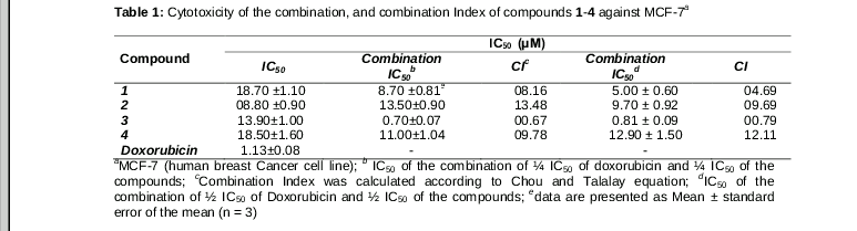
The antiproliferative activity of factors 1-4 were evaluated against the MCF-7 cell line and the results showed IC50 values of 18.70 ±1.10, 8.80 ± 0.90, 13.9 ± 1.00 and 18.5± 1.60 mg/ml, respectively.
Cell cycle distribution
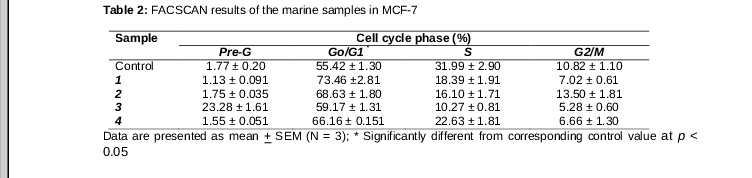
The study of the possible mode action of the antiproliferative effects of 1, 2, 3, and 4, thus, the cell cycle analysis was evaluated (Figures 2 and 3).All compounds decreased the population in S phase from 31.99 ± 2.90 to 18.39 ± 1.91, 16.10 ± 1.71, 10.27 ± 0.81 and 22.63 ± 1.81 %. Moreover, they induced a compensatory increase in the population in the fraction G0/G1 (non-proliferating cells) from 55.42 ± 1.30 to 73.46 ± 2.81, 68.63 ± 1.80, 59.17 ± 1.31 and 66.16 ± 0.151 %. All compounds induced compensatory decreased the population of MCF-7 in G2/M phase from 10.82 ± 1.10 to 7.02 ± 0.61, 13.50 ± 1.81, 5.28 ± 0.60 and 6.66 ± 1.30 %, respectively. Finally, compound 3, increased the accumulation of MCF-7 population in the pre-G phase by 23.28 ± 1.61 % which indicates its apoptotic effect.
Discussion
Compounds 1-4 exhibited cytotoxic activity against MCF-7 cells with IC50 ≤ 20 µM. These compounds have been estimated by the flow cytometry assay aimed at evaluating their effects on the cell cycle of MCF-7 cells. The CI (combination index) was evaluated to discover the possible synergistic potential, thus, decreasing the doxorubicin concentration and side effects.
The capability of inducting of intercellular apoptosis could be the main reason of the observed antiproliferative activity of the tested compounds. Thus, the combination index () was used to calculate the synergistic potential. This was performed by incubating the reagents together with the equimolar concentration of 1/4 and 1/2 of the IC50 concentration of both doxorubicin and compounds 1-4. The IC50 values of all combinations had increases than those obtained from either DOX alone or the compounds 1-4alone, except for compound 3, that showed decreases in the IC50to be0.70±0.07 and 0.81 ± 0.09 µM (). While the IC50of the combinations, DOX and compounds 1, 2, and 4 have been increased. Compounds 1, 2, and 4 showed synergistic activity when combined with the doxorubicin in the range 4.69 to 13.48, which indicate the antagonistic effects. Compound 3 showed synergistic activity when combined with the doxorubicin with combination indices (IC) values, 0.67 and 0.79 which fully agree with the Chou and Talalay value for the synergisticeffect of ≤ 0.8.
Conclusion
Chemical investigation of Red Sea marine animal S. glaucum, led to the isolation of four compounds belonging to the mevalontes (1-4). All compounds showed cytotoxic activity with IC50 in the range 8.80 ± 0.90 to 18.70 ± 1.10 µM against breast cancer cell lines (MCF-7). The antiproliferative activity of the test compounds can be ascribed to their ability to induce intercellular apoptosis. Compound 3 produced a synergistic effect with doxorubicin while the effects of compounds 1, 2 and 4 were antagonistic.
Declarations
Acknowledgement
References
Archives


News Updates